OUR PHILOSOPHY
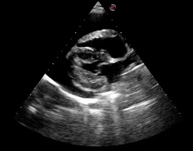
ECHOCARDIOGRAPHY
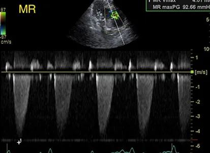
Ultrasound can be used to determine the source and extent of cardiac pathology including:
- Valve structure & motion
- Chamber sizes
- Wall thicknesses
- Myocardial contractility & heart rate
- Intra-cardiac pressures and indirect systemic and pulmonary blood pressures
- Outflow tract velocities
- Congenital defects can be accurately assessed to determine treatment options and prognosis.
- Systemic blood pressure obtained on all echocardiograms with a petMAP Graphic.
- Therapeutic options are better developed as a result of knowing the extent of the pathology present.
- Response to therapy can then be measured using the animal as his own control.
- Many cases can benefit most from referral to a cardiologist for therapeutic management or surgical intervention and the exam provided by VSI can determine when to refer the client.
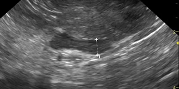
ULTRASOUND-GUIDED FLUID CAVITY CENTESIS
- Pericardial effusion
- Pleural effusion
- Ascites
- Cystocentesis – agitated collection technique for urinalysis & MIC
- Gallbladder bile culture
SOFT TISSUES
When based on ultrasound findings, the practitioner can be confident that more aggressive (and often necessary) procedures such as exploratory laparotomy are definitively indicated to further characterize or resolve a given problem.
- Abdominal exam – liver, gallbladder & biliary system, stomach, pancreas (especially now in cats), spleen, kidneys, adrenals, bladder, prostate, uterus, ovaries, bowel, vasculature, lymph nodes (mesenteric, hepatic, iliac), mesentery & serosal surfaces.
- All abdominal ultrasound exams are complete exams. It is dangerous practice to only examine part of the abdomen – all organs must be examined every time to ensure there is not occult pathology.
- Thoracic exam – if a mass effect exists on thoracic radiography, and the lesion is along the thoracic wall or the lesion is suspended in pleural effusion, the lesion may be found with ultrasound and if it can be seen, in most cases it can be biopsied to yield a diagnosis or case direction.
- Pregnancy - confirm pregnancy, estimate litter size, confirm fetal viability.
- Parathyroids & Thyroid Glands – indicated if there is hypercalcemia and elevated serum PTH. Sedation generally required providing complete relaxation of the head.
- Cryptorchid Exam – localize the cryptorchid testicle prior to surgery. Can minimize surgical time and surgeon frustration.
- Ocular Exam – used to rule-out primary neoplasia of the eye or retrobulbar space, retrobulbar abscess, retinal detatchment, retrobulbar biopsy.
ULTRASOUND-GUIDED BIOPSY
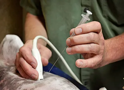
The advanced diagnostic technique of ultrasound has become a standard of care in veterinary medicine allowing the practitioner the opportunity to opt for less invasive testing. The speed and accuracy of results frequently outweigh the client & patient stress, expense, and pain of more invasive diagnostics and surgery. When combined with various minimally-invasive biopsy techniques, a diagnosis can be safely and accurately determined in most cases. Conversely when based on ultrasound findings, the practitioner can be confident that more aggressive (and often necessary) procedures such as exploratory laparotomy are definitively indicated to further characterize or resolve a given problem.
- Tru-cut tissue core biopsies (14, 16 & 18 ga.) – best for obtaining tissue architecture. Clotting profile should be performed prior and the patient will need heavy sedation or anesthesia (e.g. injectable Propofol or Medetomidine + Torbugesic is sufficient).
- Fine-needle biopsy (FNA) – the least invasive (typically non-aspiration) biopsy technique performed with special 3" needles. Generally, a normal platelet count is recommended. No clotting profile or anesthesia is necessary to obtain samples for cytopathology in the abdomen or thorax. Many disease types will yield diagnostic samples by fine-needle biopsy alone. Unless the hospital has a preferred pathologist, samples are sent by VSI to Claudia L. Barton, DVM, Diplomate ACVIM (Oncology, Internal Medicine).